-
Contents
-
Table of Contents
-
Troubleshooting
-
Bookmarks
Quick Links
Related Manuals for Zeiss Axiovert 40
Summary of Contents for Zeiss Axiovert 40
-
Page 4
Violations will entail an obligation to pay compensation. The manual is not covered by an update service. Issued by: Carl Zeiss Light Microscopy P.O.B. 4041 D-37030 Göttingen… -
Page 5
CONTENTS DESCRIPTION… -
Page 6: Operation
OPERATION…
-
Page 7: Maintenance And Troubleshooting
MAINTENANCE AND TROUBLESHOOTING ANNEX…
-
Page 8: List Of Illustrations
LIST OF ILLUSTRATIONS…
-
Page 10
NOTE • • •… -
Page 11: General View
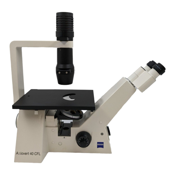
GENERAL VIEW Axiovert 40 CFL…
-
Page 13: Notes On Instrument Safety
NOTES ON INSTRUMENT SAFETY NOTE This symbol is a warning, which you must observe under all circumstances. CAUTION This symbol is a warning, which indicates a hazard to the instrument or instrument system. CAUTION This symbol is a warning, which indicates a hazard to the user of the instrument. CAUTION Hot surface! CAUTION…
-
Page 14
± … -
Page 17
Description Name and intended use Laboratory microscopes Research microscopes • • Instrument description Axiovert 40 C Axiovert 40 CFL… -
Page 18
• • • • • • • • • • • • • • • • • •… -
Page 19
1.2.1 Mechanical design Axiovert 40 C stand • • • • • • • • Axiovert 40 CFL stand • • • Fig. 1 1 Instrument models… -
Page 20
1.2.2 Optical design Fig. 1 2 Optical diagram of Axiovert 40… -
Page 21
Technical data (1) Dimensions and weight (2) Ambient conditions ° ° ° (3) Operating data… -
Page 22
± (4) Light sources (5) Optical/mechanical data… -
Page 23
³ ³ °… -
Page 24: System Overview
System overview Fig. 1 3/1 Axiovert 40 system overview…
-
Page 25
Fig. 1-3/2 Axiovert 40 system overview… -
Page 26
Fig. 1-3/3 Axiovert 40 system overview… -
Page 27
Operation Installation of the microscope 2.1.1 Unpacking • • • • • • • Fig. 2 1 Axiovert 40 packing units… -
Page 28: Installation
2.1.2 Installation (1) Preparations • • • • NOTE Fig. 2 2 Unpacking and installation (2) Screwing in of objectives • Fig. 2 3 Screwing in objectives…
-
Page 29
(3) Inserting diaphragm sliders • NOTE Fig. 2 4 Inserting diaphragm sliders (4) Mounting the HBO 50 illuminator • • • Fig. 2 5 Mounting the HBO 50 illuminator… -
Page 30
(5) Mounting the attachable object traverser M • NOTE • Fig. 2 6 Attaching the attachable object traverser M NOTE •… -
Page 31
(6) Connection to the power outlet CAUTION • • NOTE • • … CAUTION Fig. 2 7 Connection to the power outlet… -
Page 32
(7) Inserting the eyepieces • NOTE Fig. 2 8 Inserting the eyepieces (8) Compensation for defective vision 1) With one fixed and one focusing eyepiece • 2) With two focusing eyepieces • • • NOTE… -
Page 33
(9) Using the binocular tube • • (10) Inserting an eyepiece reticle (26 mm dia.) Fig. 2 9 Positions of the binocular tube • • • • • Fig. 2 10 Inserting an eyepiece reticle… -
Page 34
(11) Switching on/setting the instrument • • • • • Fig. 2 11 Switching on/setting the instrument NOTE Start-up… -
Page 35
Use of LD objectives Fig. 2 12 Working with LD objectives Table of free working distances (FWD) on Axiovert 40 Objective Name Catalogue No. with coverglass cap without coverglass cap (000000-1016-757) for bot for bottom thickness tom thickness 0.17 — 0.6… -
Page 36
Use of objectives with correction mount (corr objectives) CAUTION Use of immersion objectives CAUTION… -
Page 37
Illumination and contrast techniques • • • 2.4.1 Use of condensers • • NOTE • NOTE… -
Page 38
Height adjustment of the condenser 0.55 Á • • Fig. 2 13 Condenser 0.55 2.4.2 Brightfield illumination • • • • NOTE • Fig. 2 14 Various modes of brightfield illumination… -
Page 39: Phase Contrast
2.4.3 Phase contrast • NOTE • • • • Fig. 2 15 Observation in phase contrast Fig. 2 16 Illustration of phase-contrast adjustment • •…
-
Page 40
NOTE • • • Fig. 2 17 Inserting the annular diaphragms Objective Annular diaphragms for condenser 0.55 condenser 0.4 condenser 0.2 •… -
Page 41
2.4.4 VAREL • • • • • • NOTE Fig. 2 18 Observation with VAREL ° Fig. 2 19 VAREL with microtiter plates NOTE • •… -
Page 42
• • Objective VAREL Oblique brightfield Unilateral darkfield illumination illumination phase ring VAREL ring VAREL diaphragm Fig. 2 20 Pupil images with VAREL Preparing the slider Var, H, Var for phase contrast • • • Fig. 2 21 Slider Var, H, Var Objective VAREL diaphragms for condenser 0.55… -
Page 43
2.4.5 PlasDIC • • • • • • Fig. 2 22 Observation with PlasDIC… -
Page 44
• • • • NOTE Preparing the slider Ph, H, PlasDIC • • • NOTE Fig. 2 23 Slider Ph, H, PlasDIC… -
Page 45: Incident-Light Fluorescence
2.4.6 Incident-light fluorescence • • • • • • Fig. 2 24 Observation in incident-light fluorescence…
-
Page 46
Working with attachable object traverser M and mounting frames • • • Fig. 2 25 Working with attachable object traverser M ∅ • • • • • • • •… -
Page 47
Counting, measuring and framing reticles Crossline micrometer 14:140, d=26 Net micrometer 25/2×2, d = 26 mm ≤ Net micrometer 12.5×12.5/5;10 / d = 26 mm Framing reticle MC 2.5x / d = 26 mm Stage micrometer, positive, 5+100/100y d = 0.17 mm «… -
Page 48
Working with micromanipulators • • • Fig. 2 27 Mounting facilities for micromanipulators… -
Page 49
Replacing the specimen stage • • • Fig. 2 28 Replacing the specimen stage… -
Page 50
2.8.1 Specimen stage glass Installation • • • Fig. 2 29 Mounting the specimen stage glass 2.8.2 Heatable stage and temperable microscope stage • • • •… -
Page 51
Documentation Mounting compact digital cameras • • • • • Fig. 2 30 Camera system… -
Page 52
• • • Notes on the adjustment of the digital camera adapter D 40 M37/52×0.75 and the camera settings • • ∞ • • • Fig. 2 31 Adjusting the digital camera adapter • • • • •… -
Page 53
Maintenance and troubleshooting Maintenance • • • • ° • • • • NOTE • ° ° • •… -
Page 54
Troubleshooting and service • • (1) Checking the line voltage • • Fig. 3 1 Checking the line voltage… -
Page 55
(2) Replacing the halogen bulb CAUTION Make sure to let them cool down sufficiently! • • • • • CAUTION before • Fig. 3 2 Replacing the halogen bulb… -
Page 56
(3) Replacing the HBO 50 • • • • Wavelength in nm Fig. 3 3 HBO 50 illuminator with line spectrum… -
Page 57
Safety notes for the use of the HBO 50 CAUTION • • • CAUTION Caution: Hot surface! Replacing the HBO 50 lamp CAUTION • • • • • Fig. 3 4 Replacing the lamp of the HBO 50… -
Page 58
• CAUTION • • • • • CAUTION • • •… -
Page 59
Adjustment of the HBO 50 CAUTION • • • • Fig. 3 5 Adjustment of HBO 50 • • • Fig. 3 6 Lamp image… -
Page 60
(4) Replacing reflector modules FL P&C on 3-position reflector slider P&C • • • • • Fig. 3 7 Replacing the reflector modules FL P&C… -
Page 61
(5) Changing the filter set CAUTION • • • • Fig. 3 8 Changing the fluorescence filter set Fig. 3 9 Inserting filters and dichroic beam splitter… -
Page 62
• • (6) Changing the dichroic beam splitter CAUTION Fig. 3 10 Changing the dichroic beam splitter • • emission excitation • excitation emis sion • • Fig. 3 11 Mounting the dichroic beam splitter… -
Page 63
NOTE • excitation emis sion • • Fig. 3 12 Marking of dichroic beam splitter •… -
Page 64
(7) Service authorized… -
Page 65
ANNEX • •… -
Page 67
Index… -
Page 71
List of abbreviations…
-
Contents
-
Table of Contents
-
Troubleshooting
-
Bookmarks
Quick Links
Related Manuals for Zeiss Axiovert 40
Summary of Contents for Zeiss Axiovert 40
-
Page 4
Violations will entail an obligation to pay compensation. The manual is not covered by an update service. Issued by: Carl Zeiss Light Microscopy P.O.B. 4041 D-37030 Göttingen… -
Page 5
CONTENTS DESCRIPTION… -
Page 6: Operation
OPERATION…
-
Page 7: Maintenance And Troubleshooting
MAINTENANCE AND TROUBLESHOOTING ANNEX…
-
Page 8: List Of Illustrations
LIST OF ILLUSTRATIONS…
-
Page 10
NOTE • • •… -
Page 11: General View
GENERAL VIEW Axiovert 40 CFL…
-
Page 13: Notes On Instrument Safety
NOTES ON INSTRUMENT SAFETY NOTE This symbol is a warning, which you must observe under all circumstances. CAUTION This symbol is a warning, which indicates a hazard to the instrument or instrument system. CAUTION This symbol is a warning, which indicates a hazard to the user of the instrument. CAUTION Hot surface! CAUTION…
-
Page 14
± … -
Page 17
Description Name and intended use Laboratory microscopes Research microscopes • • Instrument description Axiovert 40 C Axiovert 40 CFL… -
Page 18
• • • • • • • • • • • • • • • • • •… -
Page 19
1.2.1 Mechanical design Axiovert 40 C stand • • • • • • • • Axiovert 40 CFL stand • • • Fig. 1 1 Instrument models… -
Page 20
1.2.2 Optical design Fig. 1 2 Optical diagram of Axiovert 40… -
Page 21
Technical data (1) Dimensions and weight (2) Ambient conditions ° ° ° (3) Operating data… -
Page 22
± (4) Light sources (5) Optical/mechanical data… -
Page 23
³ ³ °… -
Page 24: System Overview
System overview Fig. 1 3/1 Axiovert 40 system overview…
-
Page 25
Fig. 1-3/2 Axiovert 40 system overview… -
Page 26
Fig. 1-3/3 Axiovert 40 system overview… -
Page 27
Operation Installation of the microscope 2.1.1 Unpacking • • • • • • • Fig. 2 1 Axiovert 40 packing units… -
Page 28: Installation
2.1.2 Installation (1) Preparations • • • • NOTE Fig. 2 2 Unpacking and installation (2) Screwing in of objectives • Fig. 2 3 Screwing in objectives…
-
Page 29
(3) Inserting diaphragm sliders • NOTE Fig. 2 4 Inserting diaphragm sliders (4) Mounting the HBO 50 illuminator • • • Fig. 2 5 Mounting the HBO 50 illuminator… -
Page 30
(5) Mounting the attachable object traverser M • NOTE • Fig. 2 6 Attaching the attachable object traverser M NOTE •… -
Page 31
(6) Connection to the power outlet CAUTION • • NOTE • • … CAUTION Fig. 2 7 Connection to the power outlet… -
Page 32
(7) Inserting the eyepieces • NOTE Fig. 2 8 Inserting the eyepieces (8) Compensation for defective vision 1) With one fixed and one focusing eyepiece • 2) With two focusing eyepieces • • • NOTE… -
Page 33
(9) Using the binocular tube • • (10) Inserting an eyepiece reticle (26 mm dia.) Fig. 2 9 Positions of the binocular tube • • • • • Fig. 2 10 Inserting an eyepiece reticle… -
Page 34
(11) Switching on/setting the instrument • • • • • Fig. 2 11 Switching on/setting the instrument NOTE Start-up… -
Page 35
Use of LD objectives Fig. 2 12 Working with LD objectives Table of free working distances (FWD) on Axiovert 40 Objective Name Catalogue No. with coverglass cap without coverglass cap (000000-1016-757) for bot for bottom thickness tom thickness 0.17 — 0.6… -
Page 36
Use of objectives with correction mount (corr objectives) CAUTION Use of immersion objectives CAUTION… -
Page 37
Illumination and contrast techniques • • • 2.4.1 Use of condensers • • NOTE • NOTE… -
Page 38
Height adjustment of the condenser 0.55 Á • • Fig. 2 13 Condenser 0.55 2.4.2 Brightfield illumination • • • • NOTE • Fig. 2 14 Various modes of brightfield illumination… -
Page 39: Phase Contrast
2.4.3 Phase contrast • NOTE • • • • Fig. 2 15 Observation in phase contrast Fig. 2 16 Illustration of phase-contrast adjustment • •…
-
Page 40
NOTE • • • Fig. 2 17 Inserting the annular diaphragms Objective Annular diaphragms for condenser 0.55 condenser 0.4 condenser 0.2 •… -
Page 41
2.4.4 VAREL • • • • • • NOTE Fig. 2 18 Observation with VAREL ° Fig. 2 19 VAREL with microtiter plates NOTE • •… -
Page 42
• • Objective VAREL Oblique brightfield Unilateral darkfield illumination illumination phase ring VAREL ring VAREL diaphragm Fig. 2 20 Pupil images with VAREL Preparing the slider Var, H, Var for phase contrast • • • Fig. 2 21 Slider Var, H, Var Objective VAREL diaphragms for condenser 0.55… -
Page 43
2.4.5 PlasDIC • • • • • • Fig. 2 22 Observation with PlasDIC… -
Page 44
• • • • NOTE Preparing the slider Ph, H, PlasDIC • • • NOTE Fig. 2 23 Slider Ph, H, PlasDIC… -
Page 45: Incident-Light Fluorescence
2.4.6 Incident-light fluorescence • • • • • • Fig. 2 24 Observation in incident-light fluorescence…
-
Page 46
Working with attachable object traverser M and mounting frames • • • Fig. 2 25 Working with attachable object traverser M ∅ • • • • • • • •… -
Page 47
Counting, measuring and framing reticles Crossline micrometer 14:140, d=26 Net micrometer 25/2×2, d = 26 mm ≤ Net micrometer 12.5×12.5/5;10 / d = 26 mm Framing reticle MC 2.5x / d = 26 mm Stage micrometer, positive, 5+100/100y d = 0.17 mm «… -
Page 48
Working with micromanipulators • • • Fig. 2 27 Mounting facilities for micromanipulators… -
Page 49
Replacing the specimen stage • • • Fig. 2 28 Replacing the specimen stage… -
Page 50
2.8.1 Specimen stage glass Installation • • • Fig. 2 29 Mounting the specimen stage glass 2.8.2 Heatable stage and temperable microscope stage • • • •… -
Page 51
Documentation Mounting compact digital cameras • • • • • Fig. 2 30 Camera system… -
Page 52
• • • Notes on the adjustment of the digital camera adapter D 40 M37/52×0.75 and the camera settings • • ∞ • • • Fig. 2 31 Adjusting the digital camera adapter • • • • •… -
Page 53
Maintenance and troubleshooting Maintenance • • • • ° • • • • NOTE • ° ° • •… -
Page 54
Troubleshooting and service • • (1) Checking the line voltage • • Fig. 3 1 Checking the line voltage… -
Page 55
(2) Replacing the halogen bulb CAUTION Make sure to let them cool down sufficiently! • • • • • CAUTION before • Fig. 3 2 Replacing the halogen bulb… -
Page 56
(3) Replacing the HBO 50 • • • • Wavelength in nm Fig. 3 3 HBO 50 illuminator with line spectrum… -
Page 57
Safety notes for the use of the HBO 50 CAUTION • • • CAUTION Caution: Hot surface! Replacing the HBO 50 lamp CAUTION • • • • • Fig. 3 4 Replacing the lamp of the HBO 50… -
Page 58
• CAUTION • • • • • CAUTION • • •… -
Page 59
Adjustment of the HBO 50 CAUTION • • • • Fig. 3 5 Adjustment of HBO 50 • • • Fig. 3 6 Lamp image… -
Page 60
(4) Replacing reflector modules FL P&C on 3-position reflector slider P&C • • • • • Fig. 3 7 Replacing the reflector modules FL P&C… -
Page 61
(5) Changing the filter set CAUTION • • • • Fig. 3 8 Changing the fluorescence filter set Fig. 3 9 Inserting filters and dichroic beam splitter… -
Page 62
• • (6) Changing the dichroic beam splitter CAUTION Fig. 3 10 Changing the dichroic beam splitter • • emission excitation • excitation emis sion • • Fig. 3 11 Mounting the dichroic beam splitter… -
Page 63
NOTE • excitation emis sion • • Fig. 3 12 Marking of dichroic beam splitter •… -
Page 64
(7) Service authorized… -
Page 65
ANNEX • •… -
Page 67
Index… -
Page 71
List of abbreviations…
Цифровая цветная видеокамера для микроскопов под разъём C-Mount с 5 Мп CCD матрицей от SONY и USB 2.0 интерфейсом. При необходимости может быть подключена в окулярную трубку микроскопа. Для этого требуется дополнительный адаптер* (*не входит в стоимость, при покупке с камерой скидка на адаптер 50%).
Цифровая камера разработанная специально для работы с микроскопом. Камера превосходно работает со всеми сериями оптических микроскопов – биологическими, инструментальными, моно- и стереомикроскопами. Изображение наблюдаемого объекта может быть в точности передано на экран компьютера. В комплект цифровой камеры входит программа, что позволяет делать фотографии и видео — записи, а также их последующую обработку.
Цифровая окулярная камера ToupCam UHCCD05100KPA с интерфейсом USB 2.0 совместима со многими видами микроскопов. Данная модель применима в самом широком диапазоне исследований, а именно: медицина, микроскопия, наблюдение за технологическими и испытательными процессами, образовательная и научно-исследовательская деятельность.
AXIOVERT 40MAT.pdf — Focus Precision Instruments
AXIOVERT 40MAT.pdf — Focus Precision Instruments
SHOW MORE
SHOW LESS
- TAGS
- axiovert
- materials
- objectives
- zeiss
- microscope
- contrasting
- reflected
- contrast
- epiplan
- brightfield
- precision
- instruments
- www.focuspi.com

focuspi.com
You also want an ePaper? Increase the reach of your titles
YUMPU automatically turns print PDFs into web optimized ePapers that Google loves.
-
More documents
-
Similar magazines
-
Info
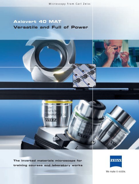
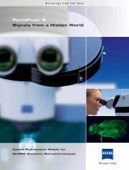
Microscopy from Carl Zeiss Axiovert 40 MAT Versatile and Full of Power The inverted materials microscope for training courses and laboratory works We make it visible.
- Page 2: In the Lead. The Flexible Solution
- Page 5 and 6: Axiovert 40 MAT Thanks to the 3-pos
- Page 7 and 8: Axiovert 40 MAT Axiovert 40 MAT. Al
- Page 9 and 10: Axiovert 40 MAT Cameras. Keeping an
- Page 12 and 13: Axiovert 40 MAT 12
- Page 14: Convincing Arguments. Your Benefits
Delete template?
Are you sure you want to delete your template?
Save as template?
Title
Description
no error
![]()
products
- FREE
- adFREE
- WEBKiosk
- APPKiosk
- PROKiosk
Resources
- Blog
- API
- Help & Support
- Status
- tuxbrain.com
- ooomacros.org
- nubuntu.org
Company
- Contact us
- Careers
- Terms of service
- Privacy policy
- Cookie policy
- Cookie settings
- Imprint
Terms of service
Privacy policy
Cookie policy
Imprint
![]() Change language
Change language
Made with love in Switzerland
© 2023 Yumpu.com all rights reserved
Назначение
Описание
Технические характеристики
Знак утверждения типа
Поверка
Сведения о методах измерений
Рекомендации к применению
Назначение
Микроскоп инвертированный Axiovert 200MAT (далее — Микроскоп) предназначен для измерений при проведении металлографических наблюдений и исследований, в том числе при контроле качества образцов в машиностроении, геологии, микроэлектронике и других отраслях промышленности.
Описание
Принцип работы Микроскопа основан на бесконтактном методе измерений размера изображения контролируемого объекта.
Метод обзора — светлое или темное поле, люминесценция, поляризация, дифференциально-интерференционный контраст.
Микроскоп оснащен двумя окулярами типа W-PL 10х/23 и пятью объективами с увеличением 5х, 10х, 20х, 50х, 100х.
Микроскоп оснащен двухстрочным ЖК-дисплеем, расположенным на магнитной опоре. В верхней строке дисплея отображается позиция и увеличение объектива, в нижней строке — яркость лампы через штриховую полосу и соответствующее значение напряжения (один штрих соответствует 0,4 V). При выключенной галогенной лампе в нижней строке ЖК-дисплея появляется информация «Hal off» вместо штриховой полосы яркости лампы.
|
т^ш |
||||
|
U4 |
||||
|
и |
||||
|
5 |
f л |
1 |
РЧ |
|
|
гм |
С ] |
р |
■гч £ |
|
|
го |
V J |
Г 1/1 |
||
|
II |
||||
Рисунок 2 — Объект-микрометр
Технические характеристики
|
Диапазон измерений, мм |
0,001 — 100 |
|
Диапазон перемещений предметного столика микроскопа, не менее, мм |
|
|
— по координате X |
0 — 100 |
|
— по координате Y |
0 — 40 |
|
Размеры предметного столика, мм |
305х170 |
|
Пределы абсолютной погрешности измерений шкалы микроскопа, мкм в диапазоне измерений: |
|
|
— 100х |
± 0,6 |
|
— 5 О X |
± 1,0 |
|
— 2 О X |
± 2,5 |
|
— 10х |
± 5,0 |
|
— 5х |
± 20 |
|
Пределы отклонения общей длины шкалы микроскопа от номинальной, мкм, при увеличении: |
|
|
— 100х |
± 5 |
|
— 5 О X |
± 10 |
|
— 2 О X |
± 20 |
|
— 10х |
± 50 |
|
— 5х |
± 100 |
|
Габаритные размеры микроскопа, мм, не более |
295x805x707 |
|
Диапазон измерений шкалы объект-микрометра, мм |
5 — О 0, |
|
Цена деления шкалы объект-микрометра, мкм |
10 |
|
Пределы абсолютной погрешности шкалы объект-микрометра, мкм |
±0,2 |
|
Пределы отклонения общей длины шкалы объект-микрометра, мкм |
±2 |
|
Масса, кг, не более |
26 |
|
Микроскоп эксплуатируется в следующих климатических условиях по гр. В1 со следующими уточнениями: |
ГОСТ 52931-2008 |
|
— температура окружающего воздуха, °С |
(20 -) |
|
— верхнее значение относительной влажности при 25 °С, без конденсации влаги, % |
80 |
|
-атмосферное давление, кПа |
100-20 |
|
Электропитание Микроскопа осуществляется от сети переменного тока: — напряжением, В -частотой, Гц |
220-23 (50±1) |
Знак утверждения типа
наносится на специальную табличку на боковой панели Микроскопа методом штемпелевания и на титульный лист руководства по эксплуатации — типографским способом.
|
Обозначение |
Наименование |
Коли чество |
Заводской номер |
Примечание |
|
Axiovert 200MAT |
Микроскоп |
1 |
3821000312 |
|
|
Головка револьверная объективов |
1 |
На 5 объективов 5х, 10х, 20х, 50х, 100х |
||
|
Источник света |
2 |
галогенный HAL100; ртутный НВО100 |
||
|
Окуляр |
2 |
W-PL |
||
|
Carl Zeiss 5+100/100 mm |
Объект-микрометр |
1 |
474027 |
0 — 5,0 мм |
|
Инструкция по эксплуатации |
1 |
|||
|
ПТИ 899-ЦКЛК-145-2013 |
Работа на инвертированном микроскопе отраженного света Axiovert 200MAT. Производственнотехническая инструкция |
|||
|
МП4435-001- 05757676 |
Методика поверки |
1 |
Поверка
осуществляется по документу МП4435-001-05757676 “Микроскоп инвертированный Axiovert 200MAT. Методика поверки”, утвержденному ФГУП «СНИИМ» в октябре 2013 г.
Эталоны: Государственный вторичный эталон единицы длины диапазоне значений 0 -1000 мм ВЭТ 2-14-59, объект-микрометр отраженного света в диапазоне значений 0 — 1 мм 2 разряда.
Сведения о методах измерений
1 ПТИ 899-ЦКЛК-145-2013 “Работа на инвертированном микроскопе отраженного света Axiovert 200MAT. Производственно-техническая инструкция”;
2 Инструкция по эксплуатации.
Нормативные и технические документы, устанавливающие требования к Микроскопу инвертированному Axiovert 200MAT
1 ГОСТ 5639-82 Стали и сплавы. Методы выявления и определения величины зерна
2 ГОСТ 8.763-2011 ГСИ. Государственная поверочная схема для средств измерений длины в диапазоне от 110-9 до 50 м и длин волн в диапазоне от 0,2 до 50 мкм
Рекомендации к применению
при выполнении работ по оценке соответствия промышленной продукции и продукции других видов, а также иных объектов, установленным законодательством Российской Федерации, обязательным требованиям.
Код товара: ArtNr.: 451221-0000-000

Краткое описание
со встроенным бинокулярного тубуса 45°/23 и 100/100-переключение между визуальным наблюдением и разъем для SLR/видеоРевольвер объектив 4-кратный HD DIC M27Эпископический Настройка HD с 2-кратным фильтры задвижки d=25 мм,… Читать далее…
- Доступность:
На складе
- Описание
- Отзывы (0)
- со встроенным бинокулярного тубуса 45°/23 и 100/100-переключение между визуальным наблюдением и разъем для SLR/видео
- Револьвер объектив 4-кратный HD DIC M27
- Эпископический Настройка HD с 2-кратным фильтры задвижки d=25 мм, задвижки съемки 3D
- Встроенный источник питания стабилизированный 12V 35W, 100…240V AC/50…60Hz/100V
Теги: штатив,
микроскоп axiovert штатив,
axiovert 40 mat,
Axio Vert.A1 штативы (),
выгодно,
баллы
Цифровая цветная видеокамера для микроскопов под разъём C-Mount с 5 Мп CCD матрицей от SONY и USB 2.0 интерфейсом. При необходимости может быть подключена в окулярную трубку микроскопа. Для этого требуется дополнительный адаптер* (*не входит в стоимость, при покупке с камерой скидка на адаптер 50%).
Цифровая камера разработанная специально для работы с микроскопом. Камера превосходно работает со всеми сериями оптических микроскопов – биологическими, инструментальными, моно- и стереомикроскопами. Изображение наблюдаемого объекта может быть в точности передано на экран компьютера. В комплект цифровой камеры входит программа, что позволяет делать фотографии и видео — записи, а также их последующую обработку.
Цифровая окулярная камера ToupCam UHCCD05100KPA с интерфейсом USB 2.0 совместима со многими видами микроскопов. Данная модель применима в самом широком диапазоне исследований, а именно: медицина, микроскопия, наблюдение за технологическими и испытательными процессами, образовательная и научно-исследовательская деятельность.
09.02.2014
•
Views
Share
Embed
Flag
AXIOVERT 40MAT.pdf — Focus Precision Instruments
AXIOVERT 40MAT.pdf — Focus Precision Instruments
SHOW MORE
SHOW LESS
ePAPER READ
DOWNLOAD ePAPER
- TAGS
- axiovert
- materials
- objectives
- zeiss
- microscope
- contrasting
- reflected
- contrast
- epiplan
- brightfield
- precision
- instruments
- www.focuspi.com
focuspi.com
You also want an ePaper? Increase the reach of your titles
YUMPU automatically turns print PDFs into web optimized ePapers that Google loves.
START NOW
-
More documents
-
Recommendations
-
Info


Microscopy from Carl Zeiss Axiovert 40 MAT Versatile and Full of Power The inverted materials microscope for training courses and laboratory works We make it visible.
- Page 2: In the Lead. The Flexible Solution
- Page 5 and 6: Axiovert 40 MAT Thanks to the 3-pos
- Page 7 and 8: Axiovert 40 MAT Axiovert 40 MAT. Al
- Page 9 and 10: Axiovert 40 MAT Cameras. Keeping an
- Page 12 and 13: Axiovert 40 MAT 12
- Page 14: Convincing Arguments. Your Benefits
Delete template?
Are you sure you want to delete your template?
Save as template?
Title
Description
no error
![]()
products
- FREE
- adFREE
- WEBKiosk
- APPKiosk
- PROKiosk
Resources
- Blog
- API
- Help & Support
- Status
- tuxbrain.com
- ooomacros.org
- nubuntu.org
Company
- Contact us
- Careers
- Terms of service
- Privacy policy
- Cookie policy
- Cookie settings
- Imprint
Terms of service
Privacy policy
Cookie policy
Cookie settings
Imprint
![]() Change language
Change language
Made with love in Switzerland
© 2023 Yumpu.com all rights reserved
Download
Table of Contents
Add to my manuals
Share
URL of this page:
HTML Link:
Bookmark this page
Manual will be automatically added to «My Manuals»
Print this page
- Manuals
- Brands
- Zeiss Manuals
- Microscope
- Axiovert 40
- Operating manual
Inverted microscope
Hide thumbs
1
2
3
4
5
6
7
8
9
10
11
12
13
14
15
16
17
18
19
20
21
22
23
24
25
26
27
28
29
30
31
32
33
34
35
36
37
38
39
40
41
42
43
44
45
46
47
48
49
50
51
52
53
54
55
56
57
58
59
60
61
62
63
64
65
66
67
68
69
70
71
72
-
page
of
72/
72 -
Contents
-
Table of Contents
-
Troubleshooting
-
Bookmarks
Table of Contents
Advertisement
2
OPERATION
Table of Contents
Previous Page
Next Page
- 1
- …
- 3
- 4
- 5
- 6
- 7
- 8
- 9
- 10
Advertisement
Table of Contents
Related Manuals for Zeiss Axiovert 40
-
Microscope Zeiss Axiovert 200M Instructions For Use
(2 pages)
-
Microscope Zeiss Axiovert 5 digital Instruction Manual
Inverted microscope for routine microscopy (56 pages)
-

Microscope Zeiss AXIOSKOP User Manual
(8 pages)
-
Microscope Zeiss Axioskop Operating Manual
Routine microscope for transmitted light and incident-light fluorescence (32 pages)
-
Microscope Zeiss Axiolab Pol Operating Manual
(116 pages)
-
Microscope Zeiss Axiolab A Operating Manual
Reflected-light (76 pages)
-
Microscope Zeiss Axioplan Universal microscope Operating Instructions Manual
(33 pages)
-
Microscope Zeiss Axiolab 5 Quick Reference Manual
Upright microscope (134 pages)


-
Microscope Zeiss Axiolab 5 Operating Manual
Upright microscope for routine and entry-level research (134 pages)
-
Microscope Zeiss Axiostar plus Operating Manual
Transmitted-light microscope (102 pages)
-
Microscope Zeiss Axio Examiner Operating Manual
Research microscope (96 pages)
-
Microscope Zeiss Axio Observer Series Operating Manual
Inverted microscope (192 pages)
-
Microscope Zeiss Axio Observer Series Quick Reference Manual
Inverted microscope (122 pages)
-
Microscope Zeiss Axiocam MR Installation Reference
Mobile stand-alone color 5 megapixel microscope camera (68 pages)
-
Microscope Zeiss AxioCam MR Installation And Reference Manual
(89 pages)
-
Microscope Zeiss Axio Imager 2 Operating Manual
Upright microscope (210 pages)


Related Content for Zeiss Axiovert 40
-
348247-9013-000 Principle Of Operation
Zeiss 348247-9013-000
-
Axiovert 5 digital Operation
Zeiss Axiovert 5 digital
-
OPMI PRO magis Operation
Zeiss OPMI PRO magis
-
LSM 510 Operation In Expert Mode
Zeiss LSM 510
-
LSM 710 Operation Of Microscope
Zeiss LSM 710
-
Crossbeam 350 Safe Operation Conditions
Zeiss Crossbeam 350
-
Crossbeam 550L Safe Operation Conditions
Zeiss Crossbeam 550L
-
Axioscope 5 Operation
Zeiss Axioscope 5


-
Primovert Operation
Zeiss Primovert
-
OPMI 1FR XY Operation
Zeiss OPMI 1FR XY
-
Axiotech Operation
Zeiss Axiotech
-
Primostar 1 Prerequisites For Commissioning And Operation
Zeiss Primostar 1
-
Axio Imager Operation
Zeiss Axio Imager
-
OPMI PROergo S7 Requirements For Operation
Zeiss OPMI PROergo S7
-
Axioskop 2 Operation
Zeiss Axioskop 2
-
Stemi DV4 Operation
Zeiss Stemi DV4


